A new study shows that in utero exposure to the mother’s antiepileptic or antidepressant drugs can affect the development of the newborn’s brain networks. In the study, new mathematical methods were developed to enable future research into how commonly used drugs or other environmental conditions affect the newborn’s brain.
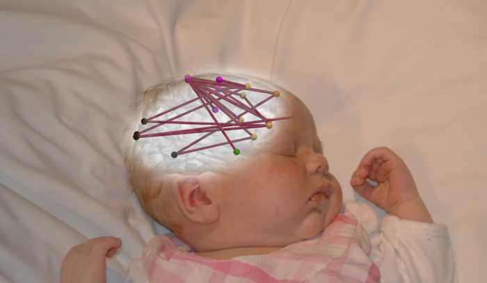
Pregnant mothers may need treatment for their medical conditions, such as mood swings or epilepsy. The effects of such drug treatment on the functions of the newborn’s brain network were examined in a study conducted at the BABA Center, a research unit at the University of Helsinki and the New Children’s Hospital of HUS Helsinki University Hospital. The study used electroencephalography (EEG) to measure the electrical activity of the brain during sleep, and the properties of the cortical network were calculated using advanced mathematical techniques.
“In previous studies, we have shown that changes in cortical activity in sleep can provide important information about the neurological condition of infants,” says senior researcher Anton Tokariev.
The study showed that exposure to antiepileptics and antidepressants during the fetal period leads to widespread changes in cortical networks, and these effects may be specific to the type of exposure to the drug. In the case of antidepressants, the effect was more pronounced in local cortical networks. In contrast, exposure to antiepileptics had drug-specific effects on the networks in the brain. Both types of drugs affect the brain networks, which are reactive to changes in sleep patterns.
“What was clinically significant in the findings was that some EEG findings were related to children’s further neuropsychological development. Stronger changes in neural networks have predicted a greater developmental deviation at the age of two, ”says Mari Videman, a pediatric neurologist at HUS Helsinki University Hospital.
It sheds new light on the early development of the brain
Studies offer a whole new way to evaluate the effects of pharmaceuticals on the development of a child’s brain function.
“The EEG measurement technique developed at the BABA Center and the state-of-the-art mathematical assessment of the brain’s associated neural networks are advances in clinical research on early neurological development,” says Professor Sampsa Vanhatalo.
Vanhatalo considers it particularly important that these EEG-based measures open a window to the mechanisms that work between neuronal cells. This leads to an opportunity to compare the results observed in human children with research using laboratory animal models. Such translational work is necessary to understand the mechanistic basis of the effects of the drug. For example, identical work is needed in animals to study how the amount or timing of treatment with maternal drugs would affect the brain function of the offspring.
“Our new methods provide a general analytical framework to support future extensive research on questions about how fetal brain development is affected by changes in the intrauterine environment. Such studies can go far beyond the mother’s drug treatment, including the mother’s nutrition and general fitness, as well as a lot of additional environmental factors, “Vanhatalo said.
COMMENTS: Tell us what you think via Twitter or Facebook Twitter